
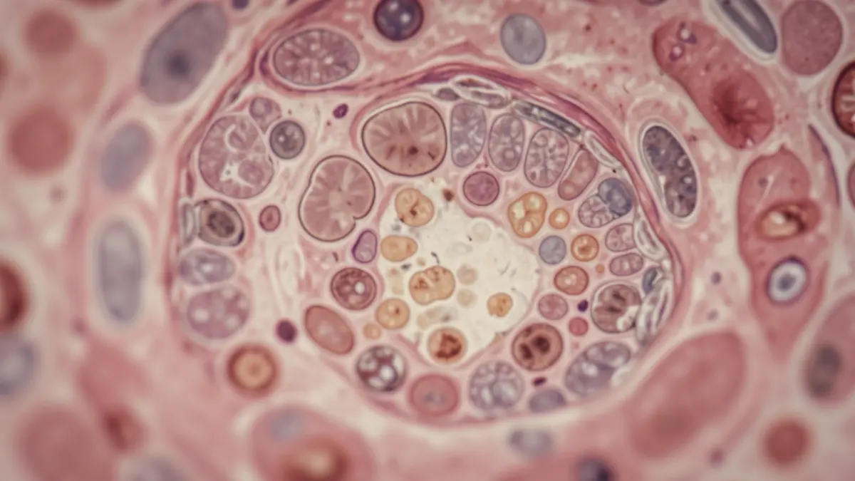
A study involving 3504 individual breast microcalcifications, scanned using high-resolution 3D micro-computed tomography (micro-CT), reveals that the surrounding tissue microenvironment offers stronger diagnostic information than the calcified core alone for distinguishing benign from malignant breast lesions. This finding by researchers from VUB-imec and UZ Brussel challenges the traditional approach focused solely on calcium deposits.
Traditionally, imaging-based assessment has focused on the MCs themselves. However, this new research, utilizing 3D micro-CT images with a spatial resolution of approximately 8 μm, analyzed 94 paraffin-embedded breast biopsy blocks. Researchers segmented 3504 individual MCs and extracted quantitative image measurements, known as radiomic features, to train machine-learning classifiers.
The results indicate that incorporating information from the MCs' surrounding tissue substantially improves classification performance. Classifiers trained exclusively on features extracted from concentric tissue shells maintained an area under the receiver operating characteristic curve (AUC) of approximately 0.81, comparable to models incorporating both the calcified core and the surrounding microenvironment. This suggests the MC-surrounding tissue is a dominant source of discriminative information.
These findings suggest that quantitative imaging approaches focusing solely on mineralized deposits may overlook valuable diagnostic information. The study, part of fundamental research using ex vivo micro-CT imaging, may guide future research into quantitative breast imaging biomarkers.